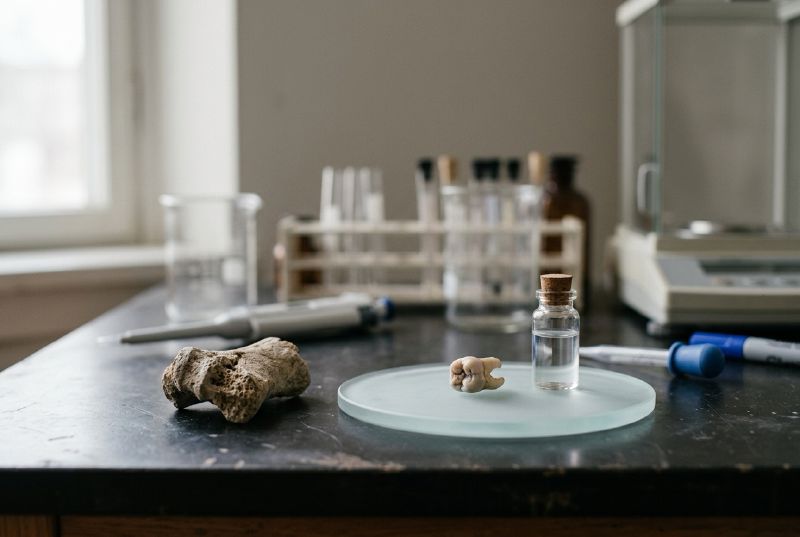
A CT scanner takes thousands of X-rays of a wrapped mummy from every angle, then a computer stacks them into a three-dimensional model a researcher can walk through, layer by layer, without touching the linen. The body stays sealed. The picture comes apart instead.
more
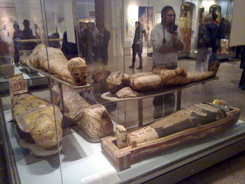
The British Museum has not unwrapped a mummy in 200 years. John Taylor, the museum’s mummy curator, said the museum wants to leave the contents of the bodies undisturbed, since they are fragile remains of people who once lived. Scanning does the job unwrapping once did, without the damage.
How the scan builds its picture
A medical CT scanner moves a rotating X-ray source and detector around a person lying still. For an object rather than a patient, the arrangement flips: the mummy sits on a turntable between a fixed X-ray source and a fixed camera, and it rotates through 360 degrees while the machine fires. Each rotation produces thousands of individual X-ray images, and software combines them into a single volumetric model.
Specialist versions push the resolution further. Micro-computed tomography, the technique a Swansea University and Egypt Centre team used on three mummified animals, produces three-dimensional images with 100 times the resolution of an ordinary hospital CT scan. Richard Johnston, the Swansea materials scientist who led that project, said the software lets him “walk around inside the body of the cat” to take fine measurements.
For a human mummy, hospital-grade dual-energy CT scanners do the work instead. The British Museum sent one of its mummies, a Theban temple singer named Tamut, through a dual-energy scanner at London’s Royal Brompton hospital. That machine let researchers make images of her blood vessels inside the wrapped body.
What the scans have found
The findings run from the routine to the startling. In Tamut’s case, the scan showed calcified plaque built up in her arteries, a warning sign a cardiologist today would treat with statins. Daniel Antoine, who curates the museum’s human remains collection, said a dislodged fragment could have caused a heart attack or a stroke. She lived roughly 2,900 years ago, and nobody could have told her that at the time.
An unidentified Theban man from around 600 BC carried a different story in his scan. Five dental abscesses would have caused him serious pain and may have contributed to his death, Antoine said. The scan also caught an embalmer’s mistake: a broken fragment of the wooden or reed spatula used to remove the brain through the nose, left behind inside the skull along with unremoved brain tissue.
Victorian-era scholars judged the pharaoh Amenhotep I’s mummy too well preserved to disturb, so it has never been unwrapped in modern times. Scans let the radiologist Sahar Saleem of Cairo University reconstruct his face and trace what happened to his body after burial. Saleem’s images showed his head had been reattached with a resin-treated linen band after his neck broke, most likely when ancient tomb robbers tore off a necklace. They also showed that priests of the 21st Dynasty had rewrapped him with care, placing gold amulets inside rather than stealing them, as some modern accounts had suggested. His right arm was folded across his chest, a pose Saleem says is the earliest known example of the style that later became standard for royal mummies. The pose was meant to echo Osiris, the god of the afterlife.
Scanning works on animals too. The Swansea team’s micro-CT scans covered a mummified cat, bird and snake. The cat was a kitten under five months old whose vertebrae separation suggests it was strangled. The snake, an Egyptian cobra, had bone fractures consistent with being killed with a whipping motion, and kidneys calcified in a pattern suggesting it had been kept with little water while alive.
Why researchers don’t unwrap them
Cutting through the wrappings destroys evidence and damages a body that, in most cases, cannot be replaced. Researchers estimate that as many as 70 million animal mummies were made as offerings for temple visitors to buy, by an established industry that bred or caught the animals.
Scanning also produces something unwrapping cannot: a model researchers can share, remeasure and reprint. The Swansea team used their cat’s scan data to 3D print a replica of its skull at twice actual size, letting them handle and measure it directly without ever touching the original.
Where the technique goes next
The technology keeps improving in resolution and in what it can distinguish. Taylor said the museum expects to keep scanning more mummies as CT technology advances. One long-term goal is to read hieroglyphic inscriptions on objects sealed inside the wrappings, such as amulets, without ever opening them.